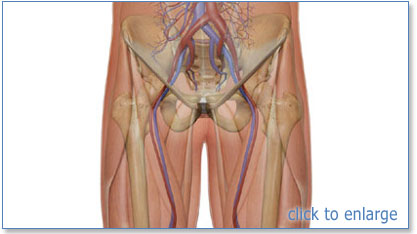
The femoral vein runs alongside the femoral artery This vein carries deoxygenated blood from your lower body, back up to your heart Anatomy Where is the femoral artery located?The femoral triangle, where the femoral artery and vein are subcutaneous, represents a site where the pulse is easily taken, where arterial blood can be sampled, and where the arterial tree can be cannulated for experimental or special diagnostic procedures Anatomy_of_femoral_artery_and_vein 2/9 Anatomy Of Femoral Artery And Vein Therapy for Peripheral Artery Disease provides a comprehensive angiographicThe nerve descends between and provides innervation to the psoas and iliacus muscle as it courses below the inguinal ligament to enter the thigh The nerve also FEMORAL NERVE • The femoral nerve is located lateral to the femoral artery
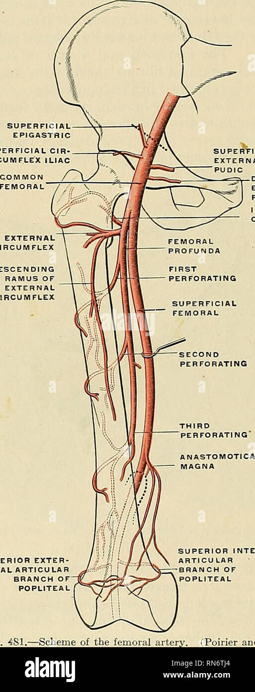
Anatomy Descriptive And Applied Anatomy 684 The Vascular Systems The Anterior Wall Of The Sheath Is A Thickened Band Of Fascia Continuous Above Poupart S Ligament With The Transversalis Fascia Called The
Femoral artery vein anatomy
Femoral artery vein anatomy- Results The modal anatomy of the femoral vein was found in 296 among 336 limbs (%) Congenital venous malformations (VM) were found in 28 of 336 limbs (12%) The results are summarized in the Table A previous anatomical study was published in 1991;Drains the legs and leads to the common iliac vein external jugular vein one of a pair of major veins located in the superficial neck region that drains blood from the more superficial portions of the head, scalp, and cranial regions, and leads to the subclavian vein femoral artery


70 Femoral Artery Stock Photos Pictures Royalty Free Images Istock
Continuation of iliac artery after passing inguinal ligament > gives off 3 superficial branches > gives off deep femoral artery branch > courses with femoral vein in adductor canal deep to sartorius m > femoral a v course through adductor hiatus > popliteal fossa Dorsalis pedis artery (DPA) The lower extremities' deep veins run adjacent to arteries of the same name which can help identify the arteries on ultrasound Figure 1 The five lower extremity arteries that are routinely examined on ultrasound include the common femoral artery (CFA), the superficial femoral artery (SFA), the popliteal artery The femoral vein ascends parallel and lateral to the great saphenous vein;
7 this study is an update with addition of colored illustrationsThese merge along with many smaller veins at the groin to form the external iliac vein Blood passing through the external iliac vein continues onward into the common iliac vein and inferior vena cava, which returns it to the heartThe femoral artery is the main provider of arterial blood supply to the thigh The femoral artery also supplies the superficial tissue of the pelvis and the anterior abdominal wall Tributaries joining the femoral artery The great saphenous vein joins the femoral vein
Knowledge of the anatomy of the common femoral artery (CFA) and common femoral vein (CFV) is important to minimize complications associated with transfemoral angiographic procedures The authors assessed variations in the relationship between the CFA and the adjacent CFV by reviewing the inguinal region of 100 computed tomographic scans of theThe femoral artery (FA) is the continuation of the external iliac artery (EIA) at the level of the inguinal ligamentAs well as supplying oxygenated blood to the lower limb, it gives off smaller branches to the anterior abdominal wall and superficial pelvis The deep femoral artery arises from the medialdorsal surface of the external iliac artery, just proximal to the pudendoepigastric trunk (D) An image of the variation c The external obturator muscle and adductor muscles are lifted up The deep femoral artery arises as a branch of the pudendoepigastric trunk (E) An image of variation d The


New Nomenclature For Femoral Vessels Journal Of Vascular Surgery


2 Femoral Vein Photos And Premium High Res Pictures Getty Images
The femoral vein is the main deep vein of the thigh and accompanies the superficial femoral artery and common femoral artery Terminology The term "superficial femoral vein" or its abbreviation, "SFV" should not be used as it is a misnomer (ie it is not a superficial vein), and can be especially confusing in the setting of deep vein thrombosisIn the human body, the femoral vein is a blood vessel that accompanies the femoral artery in the femoral sheathIt begins at the adductor hiatus (an opening in the adductor magnus muscle) and is a continuation of the popliteal veinIt ends at the inferior margin of the inguinal ligament, where it becomes the external iliac veinThe femoral vein bears valves which are mostly bicuspid and Introduction The femoral artery is a large vessel that provides oxygenated blood to lower extremity structures and in part to the anterior abdominal wall The common femoral artery arises as a continuation of the external iliac artery after it passes under the inguinal ligament The femoral artery, vein, and nerve all exist in the anterior


Femoral Artery Wikiwand
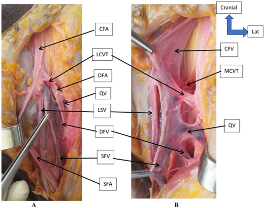


Anatomic Dissection Of The Femoral Vein At The Bamako Anatomy Laboratory
Common femoral vein on the medial side of the artery (Fig 4B) Information about the flow pattern of the vein can be assessed in the longitudinal view with Doppler US 12 Following the common femoral vein, it bifurcates into the deep femoral vein and the femoral vein (Fig 4C) In the more distal part of the medial thigh, The proximal femoral artery and vein are wrapped in a fibrous covering called the femoral sheath This sheath is made up of several components ()The lateral part of the sheath adjacent to the femoral nerve is the continuation of the iliac fascia covering the iliopsoas muscleThe femoral vein arises at the adductor canal as the continuation of the popliteal vein The femoral vein ends at the inferior margin of the inguinal ligament, becoming the external iliac vein The main tributaries of the femoral vein are the popliteal vein, the deep vein of the thigh and the great saphenous vein The femoral vein accompanies the femoral artery in the femoral sheath
:background_color(FFFFFF):format(jpeg)/images/library/13609/V4uynjLQ3qhEYyNmIqlScA_Deep_femoral_artery_01__1_.png)


Deep Femoral Artery Anatomy Branches Supply Kenhub


Lower Extremity Arterial Disease Radiology Key
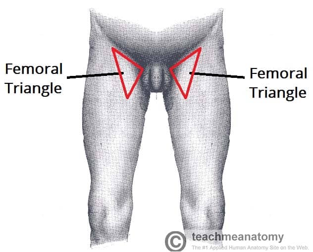
Start studying Chapter 4 Vascular Anatomy Learn vocabulary, terms, and more with flashcards, games, and other study toolsFormed when the femoral vein passes into the body cavity; Femoral Vein Anatomy continuation of the popliteal vein lies in the intermediate compartment of the femoral sheath accompanies the femoral artery in the femoral triangle at the inguinal ligament it becomes the external iliac vein FEMORAL TRIANGLE superior inguinal ligament medial border adductor longus lateral border sartorius apex sartorius crossing the adductor


Femoral Vein Wikiwand


Femoral Artery Png Images Pngwing
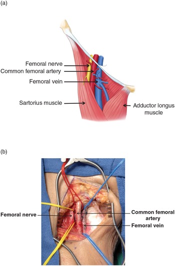
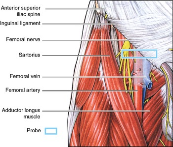
The vein is posterior to the femoral artery in the apex and medial to it at the base of the Femoral Triangle It gets the great saphenous vein and profunda femoris vein and veins corresponding to the superficial branches of femoral artery Femoral Nerve The femoral nerve is located lateral to the femoral artery, outside the femoral sheath, in This artery crosses the femoral nerve and femoral vein in such a way as to form a delta shape near the groin region This portion is known as the femoral triangle or Scarpa's triangleHere are the femoral artery and vein at the point where we saw them last, disappearing beneath the sartorius muscle To follow their course, we'll remove sartorius, and also gracilis Here's vastus medialis, here's adductor longus, with adductor magnus behind it The femoral vessels pass beneath the roof of the adductor canal, and through


Femoral Artery Wikipedia


1
Anatomy_of_femoral_artery_and_vein 2/9 Anatomy Of Femoral Artery And Vein Therapy for Peripheral Artery Disease provides a comprehensive angiographic approach to assess and determine optimal treatment strategies for peripheral artery disease (PAD) Each chapter focuses on angiography as it relates to the outcomes of endovascular workThe location of the femoral artery is at the top of your thigh in an area called the femoral triangle The triangle is just below your groin, which is the crease where The femoral artery is a continuation of the external iliac artery and constitutes the major blood supply to the lower limb In the thigh, the femoral artery passes through the femoral triangle, a wedgeshaped depression formed by muscles in the upper thighThe medial and lateral boundaries of this triangle are formed by the medial margin of adductor longus and the medial
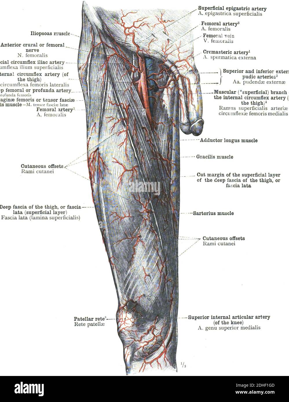


The Femoral Artery Human Anatomy Stock Photo Alamy
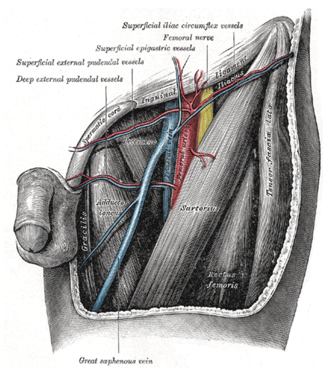


The Anatomy Of Femoral Vascular Access Taming The Sru
The femoral vein may remain on the medial side of the artery throughout its course in the thigh, or it may be doubled, especially in the adductor canal There is often a plexiform arrangement around the artery in this situation The femoral artery is the main artery that provides oxygenated blood to the tissues of the leg It passes through the deep tissues of the femoral (or thigh) region of the leg parallel to the femur The common femoral artery is the largest artery found in the femoral region of the body It begins as a continuation of the external iliac artery at The common femoral vein (CFV) lies just medial to the CFA in the proximal part of the femoral triangle, but spirals to a position posterior to the artery as they pass distally to enter the adductor canal The CFV is formed in the femoral triangle by the juncture of the superficial femoral vein (SFV) and the deep femoral vein (DFV)


Superficial Branch Of Medial Circumflex Femoral Artery Arteries Anatomy Hip Joint Anatomy


Anatomy Femoral Vein
Femoral popliteal bypass surgery is used to treat blocked femoral artery The femoral artery is the largest artery in the thigh It supplies oxygenrich blood to the leg Blockage is due to plaque buildup or atherosclerosis Atherosclerosis in the leg arteries causes peripheral vascular diseaseThe femoral artery and vein are accessible within the femoral triangle, which is defined by the inguinal ligament superiorly, the adductor longus muscle medially, and the sartorius muscle laterally The inguinal ligament is defined as a line drawn between the symphysis pubis and the anterior superior iliac spine 274 femoral vein stock photos, vectors, and illustrations are available royaltyfree See femoral vein stock video clips of 3 leg arteries leg vein anatomy circulatory system veins legs peroneal aorta illustration leg anatomy arteries veins in body leg veins lower limbs Try these curated collections
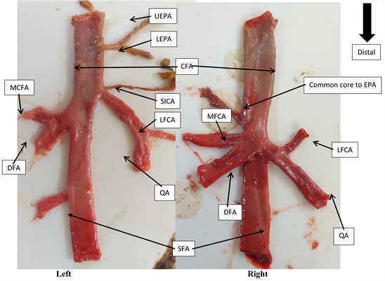


Dissection Of The Common Femoral Artery At The Bamako Anatomy Laboratory


Case Report A Rare Orientation Of Femoral Artery And Vein Sciencedirect
The femoral branch of the genitofemoral nerve is also lateral to the upper part of the femoral artery, within the femoral sheath, but lower down it passes to the front of the artery • 6 The profunda femoris artery a branch of the femoral artery itself, and its companion vein, lie behind the upper part of the femoral artery, where it lies on the pectineus – Lower down,Anatomy_of_femoral The adductor canal is a space deep to the sartorius, from the apex of the femoral triangle to the adductor hiatus The saphenous nerve passes through the adductor (Hunter's) canal along with the femoral artery and vein The nerve can become entrapped, causing the following symptoms Deep thigh ache; External Iliac Vein The external iliac vein is a continuation of the femoral vein (the major vessel draining the lower limb), arising when the femoral vein crosses underneath the inguinal ligamentIt ascends along the medial aspect of the external iliac artery, before joining with the internal iliac vein to form the common iliac vein During its short course, the external iliac vein


Medical Addicts Info The Femoral Triangle Of Scarpa The Femoral Triangle Of Scarpa Is An Anatomical Region Of The Upper Inner Human Thigh Boundaries It Is Bounded By Superiorly The Inguinal


Exposure Of The Common Femoral Artery And Vein Clinical Gate
The femoral sheath is funnelshaped and fuses with the adventitia of the vessels at the site where the greater saphenous vein joins the femoral vein4 The presence of the femoral sheath that encloses the CFA assists in preventing pseudoaneurysm formation after puncture The deep femoral artery branches 25 to 5 cm distal from the origin of the CFABrowse 954 femoral artery stock photos and images available, or search for femoral vein or vascular system to find more great stock photos and pictures plantar arteries anatomy engraving 16 femoral artery stock illustrationsStructure The femoral artery enters the thigh from behind the inguinal ligament as the continuation of the external iliac artery Here, it lies midway between the anterior superior iliac spine and the symphysis pubis Its first three or four centimetres are enclosed, with the femoral vein, in the femoral sheathIn 65% of the cases, common femoral artery lies anterior to the femoral vein


Femoral Artery Course And Branches Preview Human Anatomy Kenhub Youtube


Instant Anatomy Lower Limb Surface Femoral Artery
The surface anatomy of the femoral vein is identified for venipuncture by palpating the point of maximal pulsation of the femoral arteryKnowledge of the anatomy of the common femoral artery (CFA) and common femoral vein (CFV) is important to minimize complications associated with transfemoral angiographic procedures The authors assessed variations in the relationship between the CFA and the adjacent CFV by reviewing the inguinal reA study of the source of the blood supply to the anterolateral femoral flap was carried out on 42 lower limbs of adult cadavers (among them 35 cadavers with injection of red latex and 1 with india ink into the arteries and 6 vascular cast specimens), and the surface locations of the vascular pedicle were detected on 50 healthy adults
:watermark(/images/watermark_only.png,0,0,0):watermark(/images/logo_url.png,-10,-10,0):format(jpeg)/images/anatomy_term/femoral-artery-3/m9odBljZnOpIeioYFcTIgA_X4wsuqxApv_Arteria_femoralis_2.png)


Femoral Artery Anatomy And Branches Kenhub


70 Femoral Artery Stock Photos Pictures Royalty Free Images Istock
• The femoral artery is accompanied by the femoral vein Just below the inguinal ligament The vein is medial to the artery • However, the femoral vein gradually crosses to the lateral side posterior to the artery • Femoral vein is directly behind the artery at the apex of the femoral triangle, and lateral to the lower end of the artery
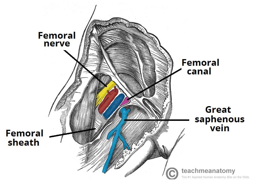


The Femoral Triangle Borders Contents Teachmeanatomy
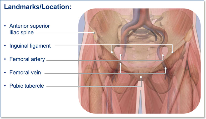


Section 2 Anatomy And Physiology
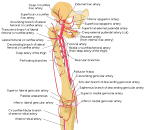


Femoral Artery Physiopedia
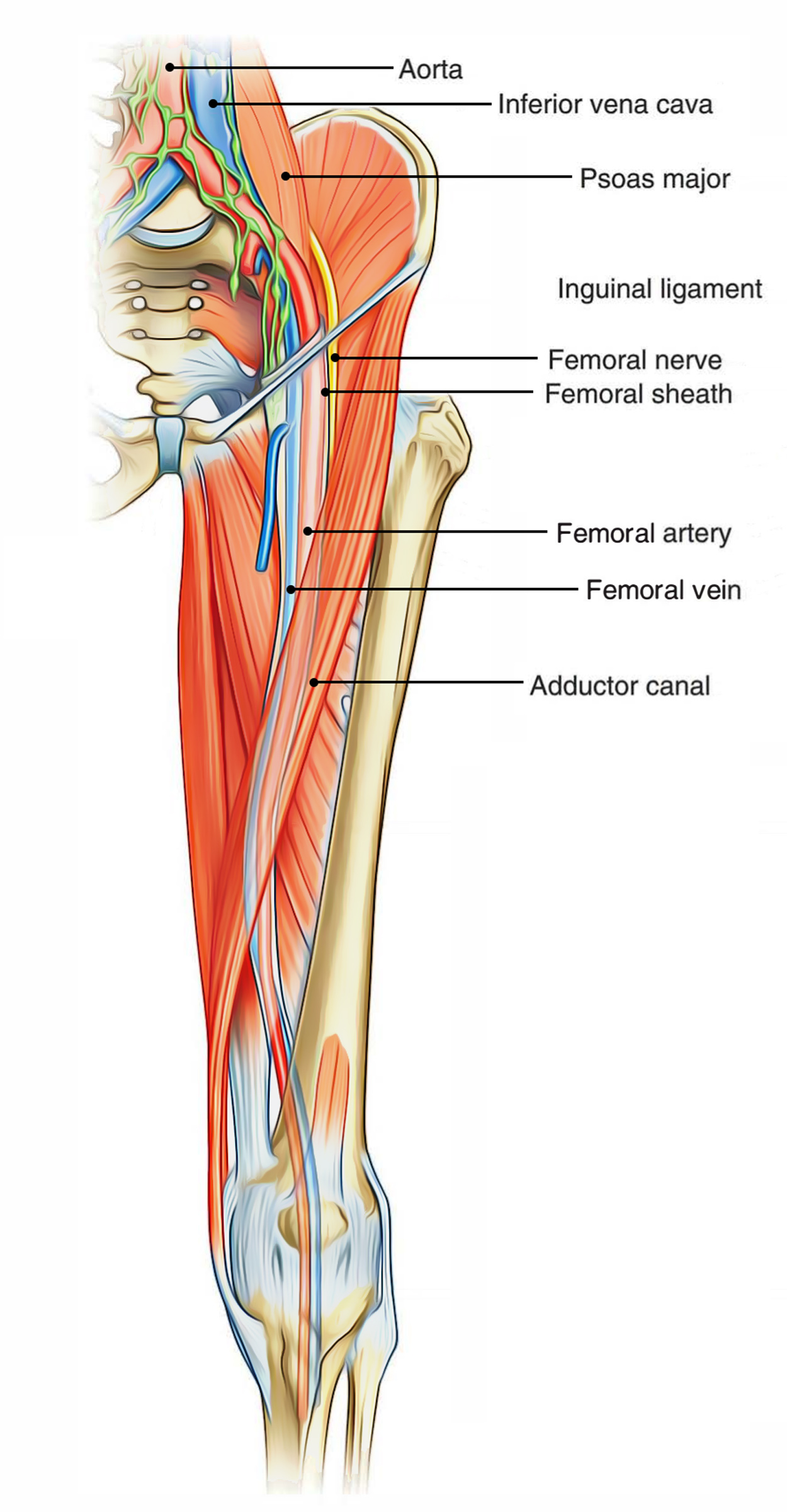


Easy Notes On Femoral Triangle Learn In Just 4 Minutes Earth S Lab


The Femoral Triangle And Superficial Veins Of The Leg Anaesthesia Intensive Care Medicine


Femoral Vein Wikipedia


Anatomy Of The Femoral Nerve Artery And Vein Medical Illustration


Femoral Vein Png Images Pngwing


Dr Francois Du Toit Department Of Diagnostic Radiology
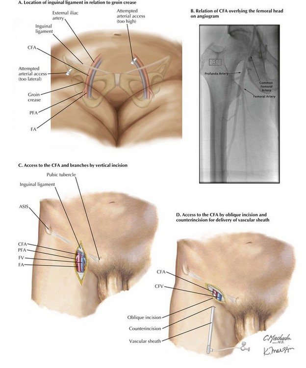


Exposure Of The Common Femoral Artery And Vein Basicmedical Key
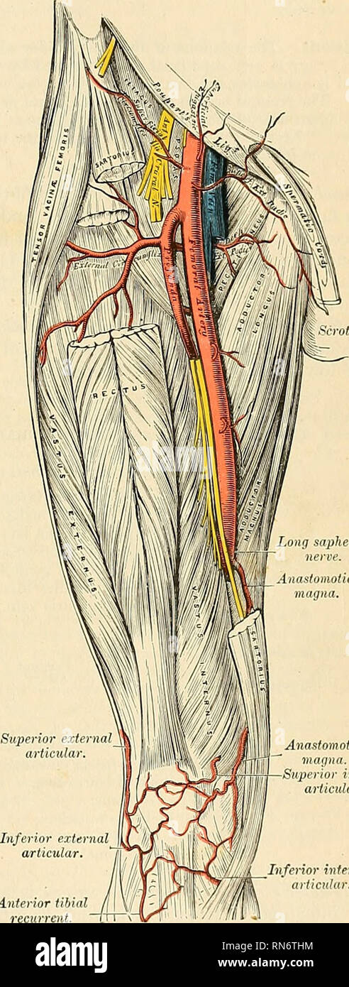


Anatomy Descriptive And Applied Anatomy The Femoral Artery G85 To Half An Inch And It Extends From The Femoral Ring To The Upper Part Of He Saphenous Opening This Canal Has


Anatomy Lectures Femoral Artery Femoral Vein Femoral Nerve Youtube
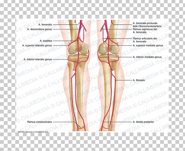


Thumb Knee Femoral Artery Popliteal Artery Crus Png Clipart Abdomen Anatomy Angle Arm Blood Vessel Free


Femoral Artery Injuries Chapter 35 Atlas Of Surgical Techniques In Trauma


Illustration Of The Femoral Nerve Block Region Showing The Femoral Download Scientific Diagram


Anatomy Descriptive And Applied Anatomy 684 The Vascular Systems The Anterior Wall Of The Sheath Is A Thickened Band Of Fascia Continuous Above Poupart S Ligament With The Transversalis Fascia Called The


Pin On Learning


Femoral Artery Femoral Nerve Mnemonics Arteries


70 Femoral Artery Stock Photos Pictures Royalty Free Images Istock


Femoral Vein Vein Vascular Center


Total Hip Replacement Doctor Stock


Femoral Artery Physiopedia


Healthy Street Anatomy Of Femoral Triangle The Femoral Facebook


Femoral Artery
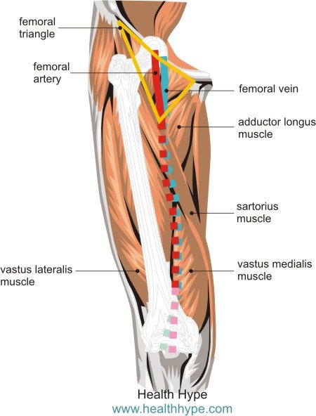


Femoral Blood Vessels Artery And Vein Anatomy Pictures Healthhype Com
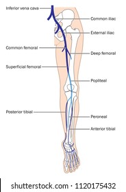


Femoral Vein Images Stock Photos Vectors Shutterstock


Femoral Artery Radiology Reference Article Radiopaedia Org


Thigh Knee And Popliteal Fossa Knowledge Amboss


Vascular Anatomy
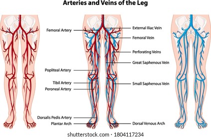


Artery Leg Images Stock Photos Vectors Shutterstock
/GettyImages-87302280-83604c7a3ca84315a84304a002377404.jpg)


Femoral Vein Anatomy Function And Significance


Femoral Vein Radiology Reference Article Radiopaedia Org


Femoral Region Gastrointestinal Medbullets Step 1


1
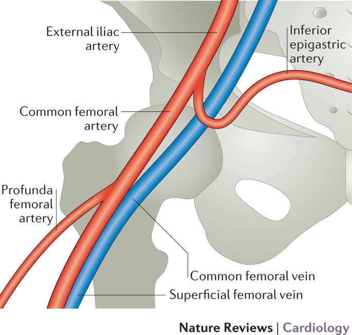


Arterial Access And Arteriotomy Site Closure Devices Nature Reviews Cardiology
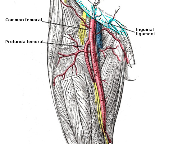


Figure Femoral Artery Anatomy Image Courtesy S Bhimji Statpearls Ncbi Bookshelf


954 Femoral Artery Photos And Premium High Res Pictures Getty Images
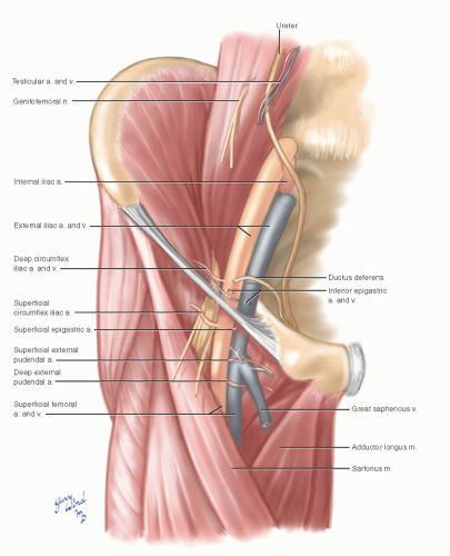


Common Femoral Artery Basicmedical Key
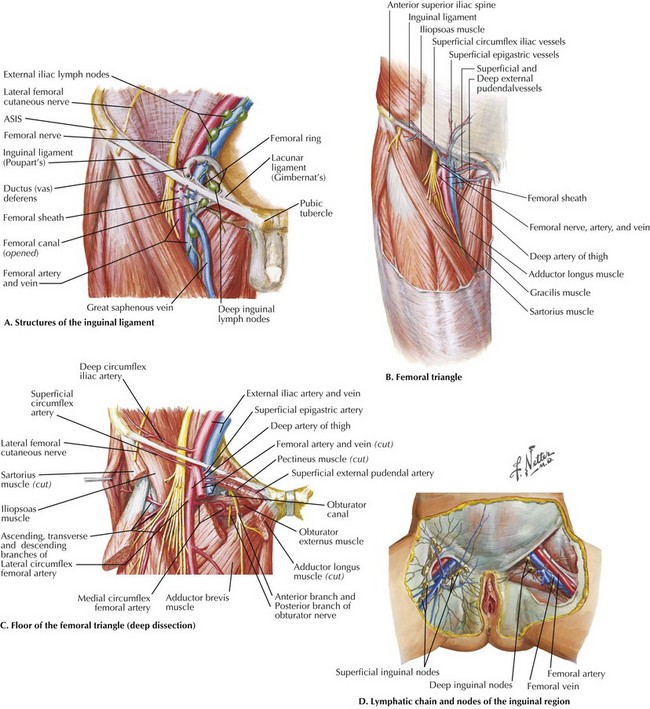


Exposure Of The Common Femoral Artery And Vein Basicmedical Key


The Femoral Triangle And Exposure Of The Femoral Artery Surgery Oxford International Edition


Femoral Artery Human Anatomy Stock Image Image Of Information Offset


The Deep Femoral Artery And Branching Variations A Case Report Arteria Profunda Femoris Ve Dallarinin Varyasyonu Olgu Sunumu Semantic Scholar


Femoral Artery Human Leg Arm Vein Arm Hand People Png Pngegg


Femoral Artery Prohealthsys


1


Femoral Artery And Vein Relevant Anatomy For Percutaneous Catheterization
:watermark(/images/watermark_only.png,0,0,0):watermark(/images/logo_url.png,-10,-10,0):format(jpeg)/images/anatomy_term/vena-iliaca-communis/H3EWTvOXMzqiwvJz3gSlw_V._iliaca_communis_01.png)


Femoral Vein Anatomy Tributaries Drainage Kenhub


The Femoral Approach Dialisis Y Trasplante
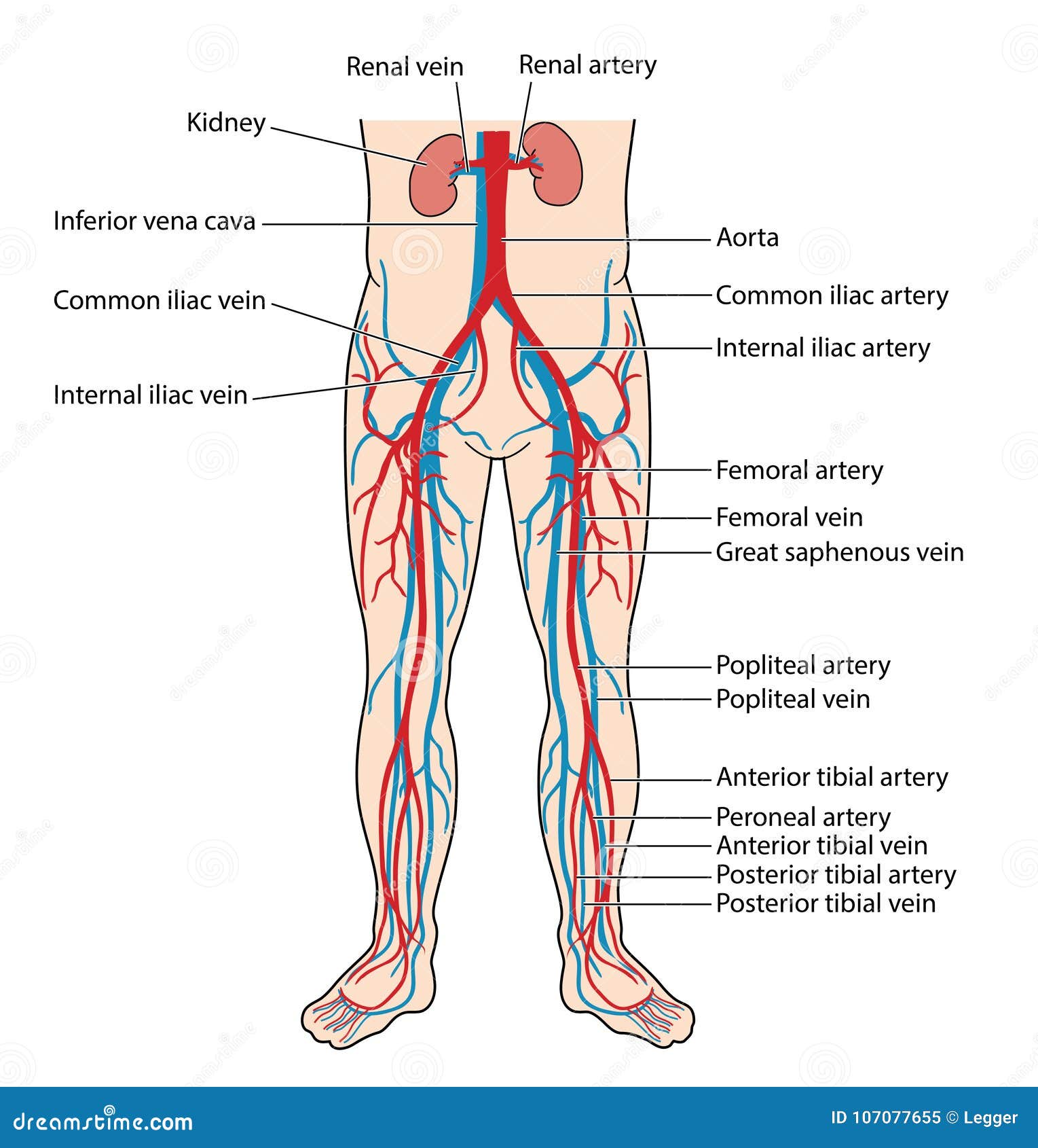


Blood Vessels Of The Lower Body Stock Vector Illustration Of Arteries Vein


Close Proximity Of The Femoral Nerve Femoral Artery And Femoral Vein To The Acetabular Retractor Trialexhibits Inc


Neurovascular Anatomy


The Anatomy Of Femoral Vascular Access Taming The Sru
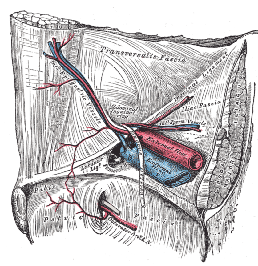


The Anatomy Of Femoral Vascular Access Taming The Sru


The Arteries Of The Lower Extremity Human Anatomy


Femoral Artery Anatomy


Human Vessels Ppt Download


Femoral Artery
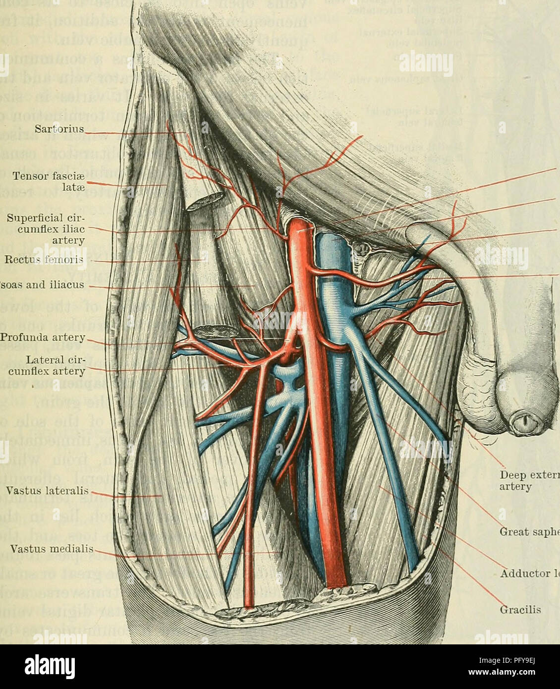


Cunningham S Text Book Of Anatomy Anatomy The Deep Veins Of The Lowek Extremity 987 The Medial Side Of The Femoral Artery About 37 Mm One And A Half Inches Below The Inguinal


Femoral Artery Anatomy Origin Course Branches And Termination Animated Lecture Youtube
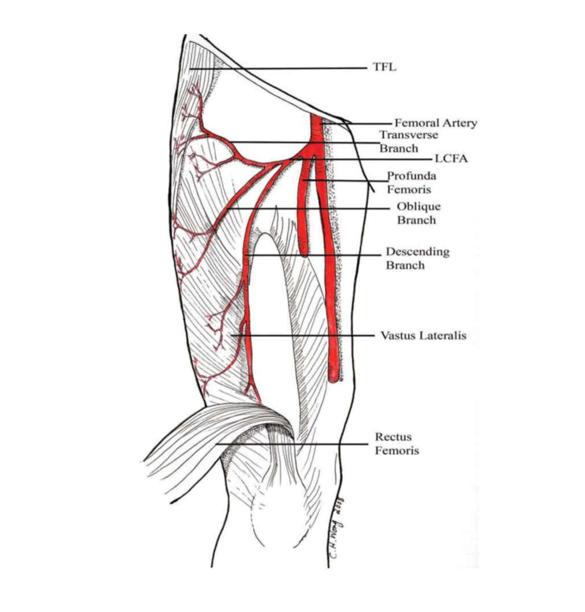


Cureus Anatomic Variations Of The Deep Femoral Artery And Its Branches Clinical Implications On Anterolateral Thigh Harvesting


Femoral Triangle Dr Bindhu S Objectives At The


The Anatomy Relevant To Percutaneous Catheterization Of The Femoral Download Scientific Diagram


The Role Of The Deep Femoral Artery As An Inflow Site For Infrainguinal Revascularization Journal Of Vascular Surgery


What Is The Femoral Vein With Pictures


Focus On Venous Embryogenesis Of The Human Lower Limbs Servier Phlebolymphologyservier Phlebolymphology


Section 2 Anatomy And Physiology


A Anatomy Of Femoral Vein B Surface Markings Of Femoral Vein Download Scientific Diagram


Approaches And Techniques For Extracorporeal Membrane Oxygenation Thoracic Key
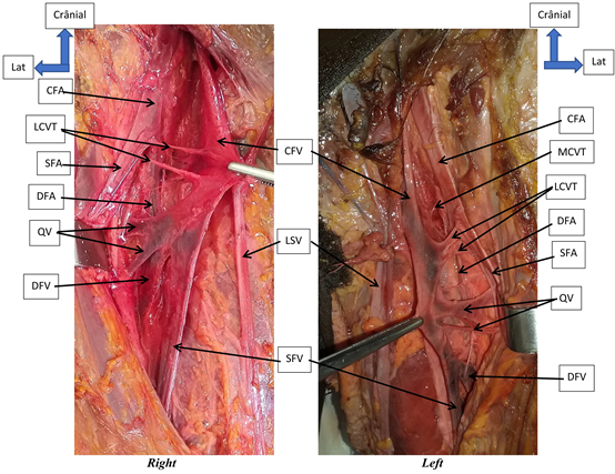


Anatomic Dissection Of The Femoral Vein At The Bamako Anatomy Laboratory


The Femoral Triangle Borders Contents Teachmeanatomy


Free Vector Arteries And Veins Of The Leg


Femoral Vein Sonoanatomy For Anaesthetists


Femoral Vein Archives Learn Muscles
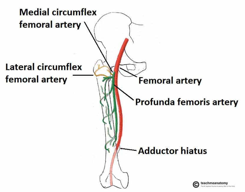


Arteries Of The Lower Limb Thigh Leg Foot Teachmeanatomy
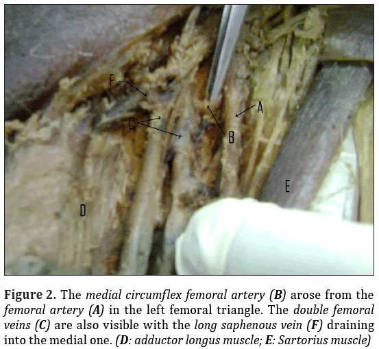


Double Femoral Veins And Other Variations In The Lower Limbs Of A Single Cadaver


Chapter 40 Femoral Artery Injuries Anesthesia Key


The Femoral Triangle And Exposure Of The Femoral Artery Surgery Oxford International Edition


1


S F Physical Exam Flashcards Quizlet
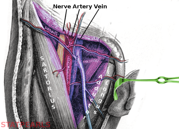


Anatomy Abdomen And Pelvis Femoral Triangle Article
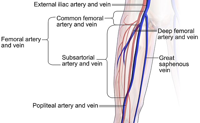


Femoral Artery Wikiwand


Untitled Document


0 件のコメント:
コメントを投稿